Retinal Photography
Retinal photography uses an advanced camera system to produce a high resolution photograph of your retina, blood vessels and optic nerve. This photograph can be analysed to screen for abnormalities assisting with early detection of diseases such as macular degeneration, glaucoma and retinal changes that may be associated with diabetes. Abnormalities that are detected can be monitored and treated to better protect the quality of your sight and overall eye health. The images are recorded by our optometrists in order to track your eye health over time and for future comparison. They help in early diagnosis and ongoing monitoring of diseases such as glaucoma, macular degeneration and diabetic retinopathy.
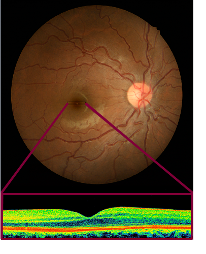
Optical Coherence Tomography (OCT) Scans
OCT scans are a painless and non-invasive way to gain a 3D view of each layer of the retina at the back of your eyes. It is a useful tool in early diagnosis and ongoing monitoring of diseases such as glaucoma, macular degeneration and other retinal conditions.
Corneal Topography
The cornea is the clear outer surface structure on the eye which receives light into the eye. Corneal topography is an imaging technology used for 3D mapping of surface curvature, which is helpful in fitting contact lenses including ortho-K contacts and in monitoring changes in corneal conditions such as keratoconus.